Endoplasmic Reticulum
Endoplasmic Reticulum (ER):
It is a complex membrane-lined network of sacs, tubules, and vesicles that runs throughout the cytoplasm of eukaryotic cells from the plasmalemma to the nuclear envelope. Procaryotic cells do not possess the same. Endoplasmic Reticulum was discovered by Porter et al (1945) and Thompson (1945) under an electron microscope. Prior to them, indications of its presence had come from the basophilic nature of some of its components (ergastoplasm of Garnier, 1897). The term endoplasmic reticulum was coined by Porter (1953). However, the organelle is not restricted to endoplasm but can extend to the plasmalemma as well as the nuclear envelope. The ER constitutes 30-60 % of the total endomembrane system of eukaryotic cells which, however, has 30-40 times the surface area as compared to that of the cell.

Occurrence:
Endoplasmic Reticulum occurs in all eukaryotic cells except mature erythrocytes. Ova, early embryonic cells, and resting cells also seem to be largely devoid of Endoplasmic Reticulum. Endoplasmic Reticulum is poorly developed in spermatocytes (as a few vesicles) and reticulocytes, simple in adipose cells (as tubules), and well developed in metabolically active cells like plasma cells, liver cells, pancreatic cells, interstitial cells of testis, etc. In muscle cells, the Endoplasmic Reticulum is known as the sarcoplasmic reticulum. Tubular and vesicular myeloid bodies of retinal cells represent components of the endoplasmic reticulum.
Structure:
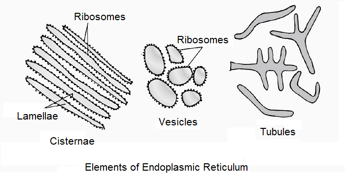
The Endoplasmic Reticulum is a network of three types of elements- cisternae, tubules, and vesicles. All of them enclose a fluid-filled space or lumen filled with an endoplasmic matrix. It is quite different from the cytoplasmic matrix. Membranes of Endoplasmic Reticulum are 50-60 Å in thickness. They are thinner than plasmalemma and most endomembranes. The Endoplasmic Reticulum is connected to the outer membrane of the nuclear envelope and perinuclear space. It may also open over plasmalemma. Connections between it and the Golgi complex have also been reported. Membranes of the Endoplasmic Reticulum possess enzymes. Enzymes present on the cytoplasmic surface include cytochrome P-450, Cyt b5, NADH—Cyt b5 reductase, NADH-Cyt c reductase, 5-nucleotidase, etc. Enzymes reported on the luminal surface are glucose 6-phosphatase, β-glucuronidase, peptidases, and nucleoside diphosphatase.
Cisternae:
They are flattened sacs of endoplasmic reticulum which generally lie parallel to one another like a stack or bundle. Interconnections occur at places. The thickness of individual cisterna is upto 40-50 nm. It is mostly due to intermembrane space filled with endoplasmic matrix. The space is wider in plasma cells, goblet cells, pancreatic cells, and others where protein synthesis is actively occurring. In such cases the endoplasmic matrix is dense. It contains abundant macromolecules and small granules.
Tubules:
They are fine tube-like extensions of the ER that form a reticular system along with cisternae and vesicles. Tubules generally occur on the periphery of the Endoplasmic Reticulum. In some cells, they also occur freely. Endoplasmic Reticulum tubules are 50-100 nm in diameter. They are both regular and irregular, unbranched, and branched. Tubules are more common in cells engaged in the storage and synthesis of lipids.
Vesicles:
Vesicles are oval or rounded sacs of the Endoplasmic Reticulum. Many of them occur free in the cytoplasm while some are found attached to other components of the Endoplasmic Reticulum. Vesicles are 25-500 nm in diameter. Pancreatic cells are rich in vesicles. In spermatocytes, the Endoplasmic Reticulum is mostly represented in the form of a few vesicles. Microsomes found during cell fractionation studies are pieces of the Plasma Membrane, Endoplasmic Reticulum, Golgi Complex, and E.R. Vesicles. Nissl granules of nerve cells and myeloid bodies of retinal cells seem to be free vesicles of the Endoplasmic Reticulum.
Types of Endoplasmic Reticulum:
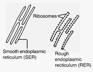
Endoplasmic Reticulum is of two main types, smooth and rough. A third type, called annulate, is also found in some cells. They may occur in continuation with each other in the same cell or as distinct structures in different types of cells.
Smooth Endoplasmic Reticulum (SER or Agranal ER):
Its membranes are smooth. They are devoid of ribophorins. Ribosomes, therefore, do not attach to their surface. SER or Agranal ER is more abundant near the plasmalemma with which it may be attached. Connections between SER and the Golgi Complex are also reported. SER is often continuous with RER. Rather SER is believed to be formed from RER. It contains fewer cisternae. The concentration of tubules and vesicles is higher. SER develops in cells engaged in the synthesis and metabolism of lipids, steroids, carbohydrates, vitamins, etc., e.g., adipose cells, interstitial cells, adrenal cortical cells, absorptive cells of the intestinal epithelium, glycogen metabolizing cells of the liver, muscle cells, retinal cells, leucocytes, etc. Sphaerosomes are believed to develop from SER. Glycosomes are special particles often found associated with fine endoplasmic tubules but actually belong to the cytoplasmic matrix. They are 50-200 nm in diameter. Enzymes associated with glycogenesis or glycogen synthesis are present in glycosomes. Membranes of SER in glycogen-storing liver cells contain enzymes involved in glycogenolysis. Myeloid bodies of retinal cells and sarcoplasmic reticulum of muscle cells are representatives of SER.
Rough Endoplasmic Reticulum (RER or Granal ER):
The membranes of RER bear a large number of ribosomes over their cytoplasmic surface. The attachment occurs by means of two types of glycoproteins, ribophorin I (65000 daltons) and ribophorin II (64000 daltons). Free ribosomes can also be associated with membranes of the Endoplasmic Reticulum by means of signal recognition particles or SRPs. An SRP is formed of a small cytoplasmic or scRNA and six polypeptides. The polypeptide formed by such ribosome contains a signal sequence at its tip which is important for association with SRP and the receptor site of the membrane. A channel is created by the receptor for the passage of synthesized polypeptide into the endoplasmic matrix. The signal sequence of the polypeptide is removed by an enzyme present on the luminal side of the membrane.
RER has more of cisternae and fewer number of tubules and vesicles. It is more abundant near the nucleus where it is connected with its outer membrane. Becuase of the presence of ribosomes, RER is basophilic (hence ergastoplasm of Garnier, 1897). Rough or Granal ER is specialized to synthesize and transport proteins. It, therefore, occurs in cells engaged in active metabolism, and secretion of proteins and enzymes, e.g., goblet cells, pancreatic acinus cells, plasma cells, fibroblasts, etc. Lysosomes are formed by the joint activity of RER and Golgi Complex. SER is involved as a bridge.
Annulate Endoplasmic Reticulum (Annulate Lamellae):
Small parts of the Endoplasmic Reticulum have been found to possess pores (Mecullo, 1952) similar to the ones present in the nuclear envelope. Structurally they can be smooth or rough. It is believed that the annulate Endoplasmic Reticulum is formed from a nuclear envelope through blebbing. During telophase, it gives rise to the nuclear envelope along with the vesicular remains of the parent nuclear envelope. Annulate Endoplasmic Reticulum has been observed in cells of invertebrates, immature oocytes, spermatocytes, and embryonic and fetal cells of vertebrates.
Biogenesis:
(i) Leskes et al (1971) believe that the Endoplasmic Reticulum always develops from the pre-existing Endoplasmic Reticulum.
(ii) Rough Endoplasmic Reticulum develops from the nuclear envelope. SER grows from RER (Seikevitz and Palade, 1966).
(iii) Endoplasmic Reticulum develops from ordinary membranes in which specific lipids, proteins, and enzymes are incorporated during differentiation.

Functions Common to SER and RER:
(1) Mechanical Support- The membranous network of the Endoplasmic Reticulum provides mechanical support to an otherwise colloidal complex of the cytoplasmic matrix.
(2) Localization of Enzymes- Endoplasmic Reticulum membranes possess sites for a number of enzymes and cytochrome to carry out specific reactions.
(3) Large Surface Area- One cubic centimetre of liver tissue possesses an Endoplasmic Reticulum of 11m2. The large surface area is useful for rapid synthesis of biochemicals.
(4) Localization of Organelles- It holds various cell organelles in their position.
(5) Desmotubules- With the help of desmotubules, the Endoplasmic Reticulum of one cell communicates with the Endoplasmic Reticulum of adjacent cells.
(6) Conduction of Information- It conducts information from outside to inside of the cell and between different organelles of the same cell.
(7) Intracellular Transport- Endoplasmic Reticulum functions as a circulating system of the cell for quick transport of materials.
(8) Vacuoles- It forms vacuoles.
(9) Nuclear Membranes- During telophase, part of the nuclear envelope is formed by the endoplasmic reticulum.
(10) Membranes to Golgi Apparatus- Endoplasmic Reticulum provides membranes to Golgi Apparatus for production of vesicles and Golgian Vacuoles.
(11) Precursors to Golgi Apparatus- It provides precursors to Golgi Apparatus for complexing and elaboration of biochemicals for internal use as well as secretion.
(12) Storage- Glycosomes or glycogen-storing particles seem to be formed by the Endoplasmic Reticulum. The Sarcoplasmic Reticulum of muscle cells stores calcium. Ca2+ ions are released at the time of muscle contraction.
(13) Detoxification (Metabolism of Xenobiotics)- Living beings are exposed to a large number of foreign substances, pollutants, carcinogens, drugs, etc. They are extremely harmful. Two cytochromes, located mostly on the smooth membranes of the Endoplasmic Reticulum (especially for liver cells) and mitochondria are known to metabolize them. They are P-450 and P-448. P-448 hydroxylates polycyclic aromatic hydrocarbons (PAAs) which are generally carcinogens. P-450 hydroxylates a number of pollutants, carcinogens, and drugs. About 50% of the drugs taken by patients are metabolized by it. Six species of P-450 have been isolated from liver ER.
Functions Specific to RER:
(1) SER- SER is formed from RER through the loss of ribosomes.
(2) Ribophorins- It possesses ribophorins for holding ribosomes over it.
(3) Protein Processing- The luminal side of RER possesses enzymes for processing polypeptides synthesized by attached ribosomes.
(4) Large Surface Area- RER has a large surface area which provides proper space to ribosomes for their activity without interference from others.
(5) Proteins for Transport- Proteins formed by ribosomes attached to RER enter its lumen for intracellular and extracellular transport.
(6) Enzymes for Lysosomes- Zymogens of lysosome enzymes are synthesized by RER.
Functions Specific to SER:
(1) Ascorbic Acid- Its synthesis occurs over SER.
(2) Retinal Pigments- They are formed from vitamin A over SER in retinal cells.
(3) Glycogenolysis- Enzymes located over SER of liver cells take part in the breakdown of glycogen.
(4) Fat Synthesis- SER takes part in fat synthesis inside adipose cells.
(5) Sphaerosomes- They are believed to be formed from SER.
(6) Steroid Hormones- SER takes part in the synthesis of steroid hormones, e.g., testosterone in the testis, and estrogens in the ovary.